
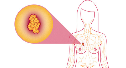
Breast cancer is the most frequently diagnosed life-threatening cancer in women. It is the leading cause of cancer death among women.
Many early breast carcinomas may be asymptomatic; pain or discomfort is not usually a symptom of breast cancer. Breast cancer is often first detected as an abnormality on a mammogram before the patient or healthcare provider feels it.

Increased public awareness and improved screening have led to earlier diagnosis, at stages amenable to complete surgical resection and curative therapies. Consequently, survival rates for breast cancer have improved significantly, particularly in younger women.
Surgery is considered primary treatment for breast cancer. Many patients with early-stage breast cancer are cured with surgery alone.
Surgery is considered primary treatment for breast cancer, as many patients with early-stage disease are cured with surgery alone. The goals of breast cancer surgery include complete resection of the primary tumor with negative margins to reduce the risk of local recurrences, and pathologic staging of the tumor and axillary lymph nodes for providing necessary prognostic information. Several different types of operations are available for the treatment of breast cancer.
Adjuvant treatment for breast cancer involves radiation therapy and a variety of chemotherapeutic and biologic agents.
Numerous prognostic and predictive factors for breast cancer have been identified by the College of American Pathologists (CAP) to guide the clinical management of women with breast cancer.
Women should be ‘breast aware’ and check their breasts regularly. Breast awareness means being familiar with your breasts and what is normal for them so you can recognise any changes easily and quickly. If you notice a lump on your breast, or any change in its appearance, feel or shape, you should have it checked by your doctor as soon as possible.
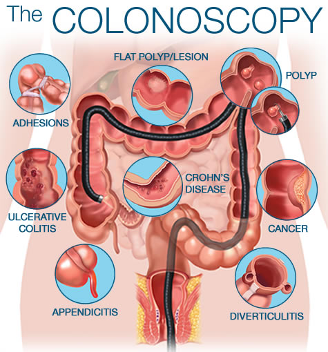
Colonoscopy is usually done as an out-patient or day case. Before your procedure you will be asked to change into a hospital gown and lie on your left side with you legs in a curled position. The procedure itself takes around 20-30 minutes. A colonoscopy does not hurt, but it can be a little uncomfortable, particularly when the scope is first passed into the anus.
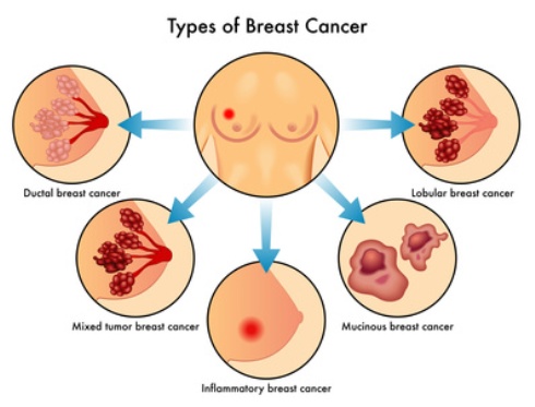
Common types of benign breast lumps include:
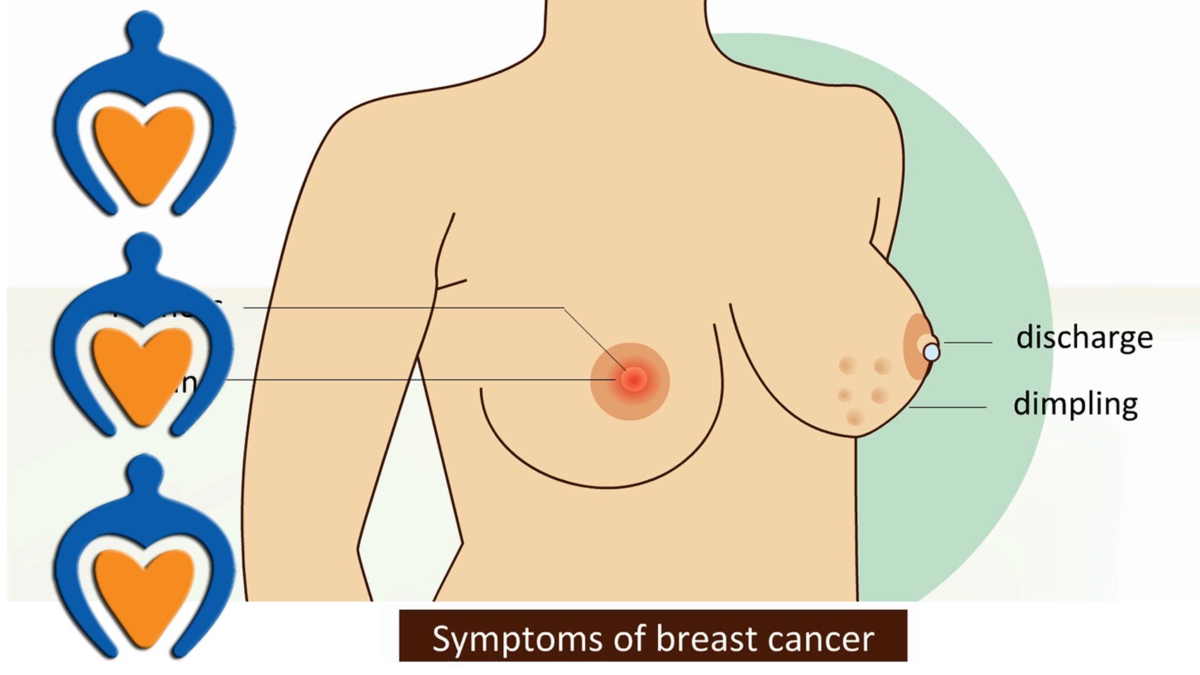
You should see your surgeon if you notice any changes to your breasts such as:
Most colonoscopies are done without any problems. If you have had a sedatve you may feel tired or sleepy for several hours afterwards. You may pass a small amount of blood from your anus if a biopsy was taken, or a polyp removed, Occasionally the colonoscope may cause damage to the colon. This may cause bleeding, infection and rarely, perforation. If any of the following occur within 48 hours after a colonoscopy, it is important that you consult your doctor immediately:
Your doctor will carry out a physical examination of both of your breasts, including the lump, if any, and may refer you for further tests. Some of the tests that you may have are explained below.
Regular breast screening is very important because it will help detect any small changes before you have other signs or symptoms. Screening is a way of identifying people who may have an increased risk of developing a certain condition. For breast cancer, screening is carried out using mammograms. This is a procedure that uses X-rays to produce an image of the inside of your breasts.
A mammogram is a simple procedure that uses X-rays to create an image of the inside of your breasts. It is used to help identify early changes in your breast tissue. Younger women usually have denser breasts than older women, which makes changes more difficult to identify. Therefore, mammograms are not as effective in women who are under 35 years of age. If you are under 35, your GP may suggest that you have a breast ultrasound instead (see below).
If you need to have a mammogram, a radiographer (an X-ray specialist) will position one of your breasts on a flat X-ray plate. A second X-ray plate will press down on your breast from above, so that it is temporarily flattened between the two plates. An X-ray will then be taken, which will produce a clear image of the inside of your breast. After the first X-ray has been taken, the same procedure will be carried out on your other breast. A mammogram only takes a few minutes to carry out. You may find it a bit uncomfortable or even slightly painful. After the procedure is complete, the mammogram (the image of your breast) will be examined for anything unusual.
may need to have a breast biopsy if the cause of your breast lump cannot be diagnosed using a mammogram or ultrasound. A biopsy is a procedure that involves removing a tissue sample from the lump for further testing. During a biopsy, a fine needle will be inserted into the lump in your breast so that a sample of tissue can be removed. The sample will then be examined under a microscope to confirm the diagnosis.
If there is any abnormality seen on the screening mammogram films, which the surgeon is not able to palpate clinically, then you are advised to have the biopsy of the suspected area done under mammographic guidance. It is computer aided exercise. The patient actually lies on the mammogram table, and the area of suspicion in the breast is 3 dimensionally targeted (like a missile on the radar operation) and then a small needle is inserted under local anaesthesia. A small amount of liquid is aspirated which is sent for cytology.
Approximately 10 to 15 percent of all breast cancers are thought to be familial and about one third of these cases are due to an inherited mutation in a BRCA1 or BRCA2 breast cancer susceptibility gene. BRCA1 and 2 mutations are associated with early-onset breast cancer, and some experts call for aggressive Screening of affected persons.
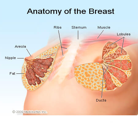
The female breast is a complex gland that is made up of several different types of tissue. Each breast contains milk glands and milk ducts for transporting milk. These structures can increase or decrease in both size and number as and when they are needed. For example, during pregnancy, the milk ducts in the breasts will grow and the breasts will get larger. During breastfeeding, the breasts may change size several times throughout the day as milk is produced and the baby feeds. The breast is made up of fibrous connective tissue, fatty tissue, nerves, blood vessels and lymph nodes (small oval-shaped glands that remove unwanted bacteria and particles from the body)
Breast lumps are quite common. About 3 in every 100 women visit their doctor regarding a problem with their breasts. Most breast lumps, about 9 out of 10, are benign (non-cancerous).There are several types of benign breast lump. They can vary in appearance or texture depending on the type. Most breast lumps are caused by hormonal changes that occur at different times in a woman’s life, such as during the menstrual cycle when a woman gets her monthly period.
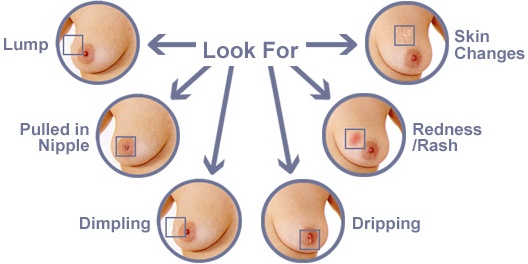
It is important to be aware of how your breasts usually look and feel so you can quickly pick up on any changes that may occur. See your doctor if you notice a lump in your breast or any change in its appearance, feel or shape.
Common types of benign breast lumps include:
Fibrocystic breast disease, also known as fibroadenosis, is a term used to describe a group of benign (non-cancerous) conditions that affect the breast. The symptoms of fibroadenosis include: • breast pain (mastalgia) • breast enlargement • lumpiness of the breast (nodularity), particularly just before or during a period Fibroadenosis can develop in one or both breasts, or can affect just part of one breast. The symptoms can also vary significantly between women, with some women finding them slightly annoying and others finding them very painful. The pain and lumpiness will usually disappear after your period. The cause of fibroadenosis is not well understood. However, it may be the result of the breast tissue responding abnormally to hormonal changes that occur with the menstrual cycle.
A fibroadenoma is a smooth, well-rounded solid lump (tumour) that sometimes develops outside the milk ducts. Milk ducts are the tiny tubes in the breast that carry milk. Fibroadenomas are made up of fibrous and glandular tissue, which has a rubber-like texture and moves easily when touched. It is also known as a “breast mouse”. A fibroadenoma will sometimes disappear, but it can remain and grow larger, particularly during pregnancy.
A breast cyst is a fluid-filled sac that develops within the breast tissue and may feel like a soft grape. Breast cysts are very common and normal. Cysts form as a natural part of the ageing of breast tissue and are most commonly found in women aged 35-50. Cysts vary in size. Some can be very tiny, while others can grow up to several centimetres in diameter. Single or multiple cysts can occur in one or both breasts. Cysts often do not cause any symptoms, although some women may experience pain, particularly if the cyst increases in size during the menstrual cycle. They do not significantly increase the risk of breast cancer developing.
A breast abscess is a painful collection of pus that forms under the skin of the breast. It can also cause a high temperature (fever) of 38°C (100.4°F) or above inflammation (redness and swelling). An abscess can also develop in a female even if she is not lactating.
While you should always see your doctor about any changes to your breasts, benign (non-cancerous) breast lumps:
Hormonal changes are the most common cause of benign breast lumps. Hormones are chemicals that are produced by the body and have a wide range of effects. Sometimes changes in the levels of hormones in your body can cause your breasts to feel lumpy or swollen.
In most cases, a benign (non-cancerous) breast lump does not need any treatment unless the lump is particularly large or painful. After diagnosing the cause, your doctor will advise you about any treatment that is necessary. If treatment is not necessary, you may be asked to return if you notice any further changes to your breasts.
Most breast lumps cannot be prevented because they are caused by hormonal changes that you have no control over. However, it is very important that you identify a breast lump as soon as it develops and get it checked by your doctor to rule our breast cancer.
Most of the time, if the lump is more than 2 cms in size, the doctor will advise the excision of the lump. It is a short procedure lasting about 30 minutes which is performed under general anesthesia. Usually the incision is taken in a cosmetic fashion in the around the areola and the lump is removed. This lump is then sent for histopathological check up which is mandatory for all cases.
If the patient has a cyst, usually the symptomatically painful cyst is aspirated in the outpatient and the fluid which is collected is sent for cytological examination. If there is rapid reaccumulation of fluid in the cyst on follow up or the patient had severe discomfort then the cyst is removed surgically and evaluated further.